

The brain enables the mind-seeing, hearing, smelling, feeling, remembering, thinking, speaking, dreaming, loving. Moreover, it is the brain that self-reflectively analyzes the brain. When we're thinking about our brain, we're thinking with our brain - by firing across millions of synapses and releasing billions of neurotransmitter molecules. Neuroscientists tell us that the mind is what the brain does. Brain, behavior, and cognition are an integrated whole. But precisely where and how are the mind's functions tied to the brain? Let's first see how scientists explore such questions.
Older brain networks sustain basic life functions and enable memory, emotions, and basic drives. Newer neural networks within the cerebrum - the hemispheres that contribute 85 percent of the brain's weight-form specialized work teams that enable our perceiving, thinking, and speaking. Like other structures above the brainstem (including the thalamus, hippocampus, and amygdala), the cerebral hemispheres come as a pair. Covering those hemispheres, like bark on a tree, is the cerebral cortex, a thin surface layer of interconnected neural cells. It is your brain's thinking crown, your body's ultimate control and information-processing center.
As we move up the ladder of animal life, the cerebral cortex expands, tight genetic controls relax, and the organism's adaptability increases. Frogs and other small-cortex amphibians operate extensively on preprogrammed genetic instructions. The larger cortex of mammals offers increased capacities for learning and thinking, enabling them to be more adaptable. What makes us distinctively human mostly arises from the complex functions of our cerebral cortex.
If you opened a human skull, exposing the brain, you would see a wrinkled organ, shaped somewhat like the meat of an oversized walnut. Without these wrinkles, a flattened cerebral cortex would require triple the area - roughly that of a large pizza. The brain's left and right hemispheres are filled mainly with axons connecting the cortex to the brain's other regions. The cerebral cortex - that thin surface layer - contains some 20 to 23 billion nerve cells and 300 trillion synaptic connections (de Courten-Myers, 2005). Being human takes a lot of nerve.
Supporting these billions of nerve cells are nine times as many spidery glial cells ("glue cells"). Neurons are like queen bees; on their own they cannot feed or sheathe themselves. Glial cells are worker bees. They provide nutrients and insulating myelin, guide neural connections, and mop up ions and neurotransmitters. Glia may also playa role in learning and thinking. By "chatting" with neurons they may participate in information transmission and memory (Fields, 2009; Miller, 2005).
In more complex animal brains, the proportion of glia to neurons increases. A postmortem analysis of Einstein's brain did not find more or larger-than-usual neurons, but it did reveal a much greater concentration of glial cells than found in an average Albert's head (Fields, 2004).
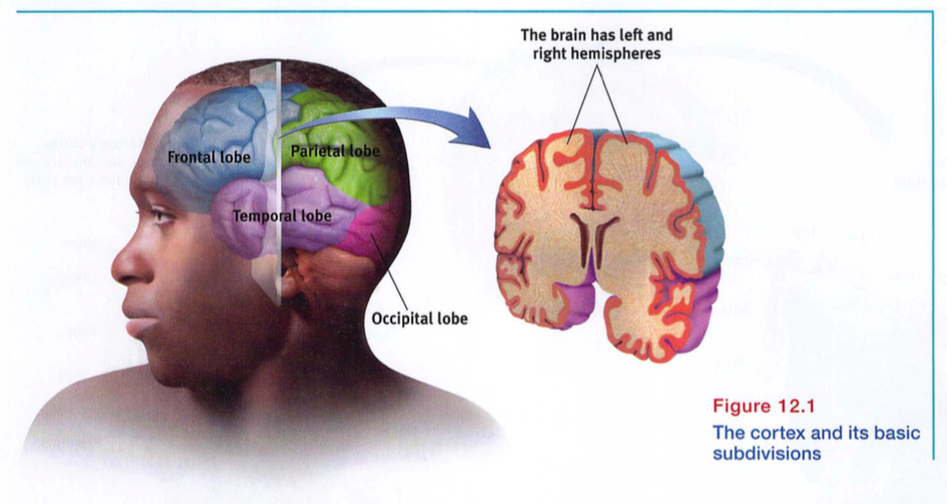
Each hemisphere's cortex is subdivided into four lobes, separated by prominent fissures, or folds (FIGURE 12.1). Starting at the front of your brain and moving over the top, there are the frontal lobes (behind your forehead), the parietal lobes (at the top and to the rear), and the occipital lobes (at the back of your head). Reversing direction and moving forward, just above your ears, you find the temporal lobes. Each of the four lobes carries out many functions, and many functions require the interplay of several lobes.
More than a century ago, surgeons found damaged cortical areas during autopsies of people who had been partially paralyzed or speechless. This rather crude evidence did not prove that specific parts of the cortex control complex functions like movement or speech. After all, if the entire cortex controlled speech and movement, damage to almost any area might produce the same effect. A TV with its power cord cut would go dead, but we would be fooling ourselves if we thought we had "localized" the picture in the cord.
Scientists had better luck in localizing simpler brain functions. For example, in 1870, German physicians Gustav Fritsch and Eduard Hitzig made aIL important discovery: Mild electrical stimulation to parts of an animal's cortex made parts of its body move. The effects were selective: Stimulation caused movement only when applied to an arch-shaped region at the back of the frontal lobe, running roughly ear-to-ear across the top of the brain. Moreover, stimulating parts of this region in the left or right hemisphere caused movements of specific body parts on the opposite side of the body. Fritsch and Hitzig had discovered what is now called the motor cortex.
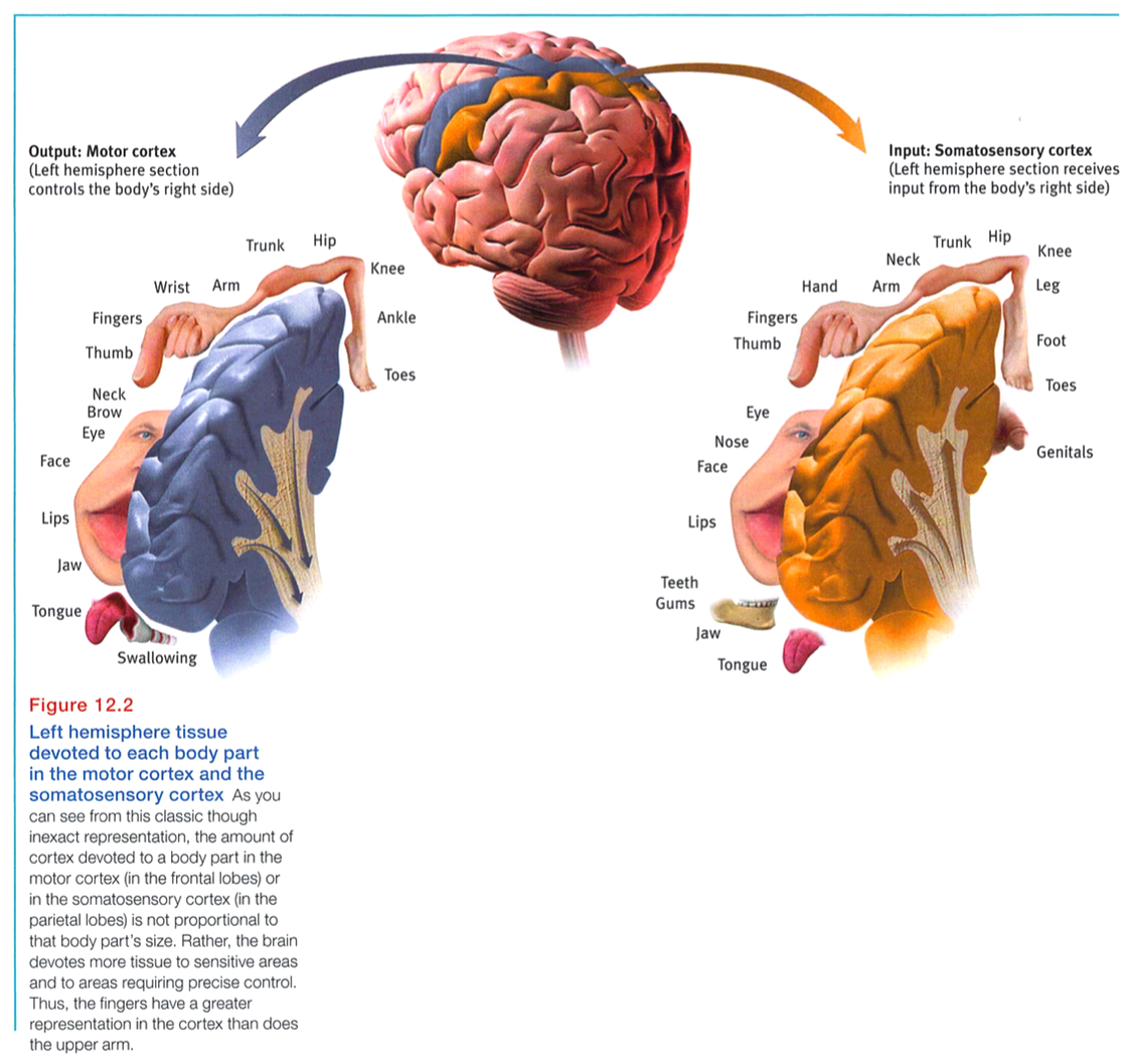
Lucky for brain surgeons and their patients, the brain has no sensory receptors. Knowing this, Otfrid Foerster and Wilder Penfield were able to map the motor cortex in hundreds of wide-awake patients by stimulating different cortical areas and observing the body's responses.
They discovered that body areas requiring precise control, such as the fingers and mouth, occupy the greatest amount of cortical space (FIGURE 12.2).
In one of his many demonstrations of motor behavior mechanics, Spanish neuroscientist Jose Delgado stimulated a spot on a patient's left motor cortex, triggering the right hand to make a fist. Asked to keep the fingers open during the next stimulation, the patient, whose fingers closed despite his best efforts, remarked, "I guess, Doctor, that your electricity is stronger than my will" (Delgado, 1969, p. 114).
More recently, scientists were able to predict a monkey's arm motion a tenth of a second before it moved-by repeatedly measuring motor cortex activity preceding specific arm movements (Gibbs, 1996). Such findings have opened the door to research on brain-controlled computers.
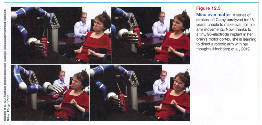
By eavesdropping on the brain, could we enable someone-perhaps a paralyzed person-to move a robotic limb? Could a brain-computer interface command a cursor to write an e-mail or search the Internet? To find out, Brown University brain researchers implanted 100 tiny recording electrodes in the motor cortexes of three monkeys (Nicolelis & Chapin, 2002; Serruya et al., 2002). As the monkeys used a joystick to move a cursor to follow a moving red target (to gain rewards), the researchers matched the brain signals with the arm movements. Then they programmed a computer to monitor the signals and operate the joystick. When a monkey merely thought about a move, the mind-reading computer moved the cursor with nearly the same proficiency as had the reward-seeking monkey. In follow-up experiments, two monkeys were trained to control a robot arm that could grasp and deliver food (Velliste et al., 2008), and then a human did the same (FIGURE 12.3).
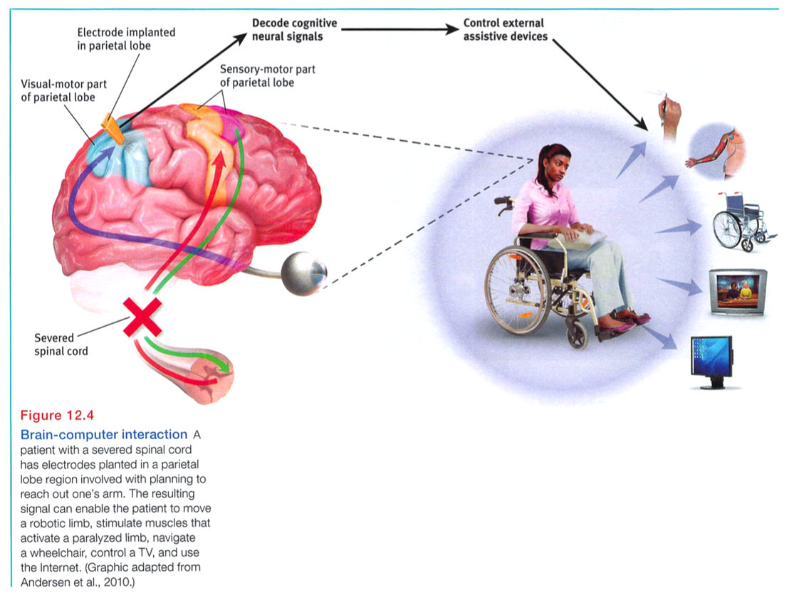
Clinical trials of such cognitive neural prosthetics are now under way with people who have suffered paralysis or amputation (Andersen et al., 2010; Nurmikko et al., 2010). The first patient, a paralyzed 25-year-old man, was able to mentally control a TV, draw shapes on a computer screen, and play video games-all thanks to an aspirin-sized chip with 100 microelectrodes recording activity in his motor cortex (Hochberg et al., 2006). If everything psychological is also biological - if, for example, every thought is also a neural event-then microelectrodes perhaps could detect thoughts well enough to enable people to control events, as suggested by FIGURE 12.4.
If the motor cortex sends messages out to the body, where does the cortex receive the incoming messages? Wilder Penfield also identified the cortical area that specializes in receiving information from the skin senses and from the movement of body parts. This area at the front of the parietal lobes, parallel to and just behind the motor cortex, we now call the somatosensory cortex (Figure 12.2). Stimulate a point on the top of this band of tissue and a person may report being touched on the shoulder; stimulate some point on the side and the person may feel something on the face.
The more sensitive the body region, the larger the somatosensory cortex area devoted to it (Figure 12.2). Your supersensitive lips project to a larger brain area than do your toes, which is one reason we kiss with our lips rather than touch toes. Rats have a large area of the brain devoted to their whisker sensations, and owls to their hearing sensations.
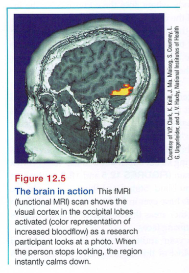
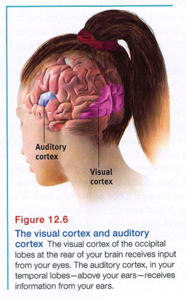
Scientists have identified additional areas where the cortex receives input from senses other than touch. At this moment, you are receiving visual information in the visual cortex in your occipital lobes, at the very back of your brain (FIGURES 12.5 and 12.6). A bad enough bash there would make you blind. Stimulated there, you might see flashes of light or dashes of color. (In a sense, we do have eyes in the back of our head!) From your occipital lobes, visual information goes to other areas that specialize in tasks such as identifying words, detecting emotions, and recognizing faces.
Any sound you now hear is processed by your auditory cortex in your temporal lobes (just above your ears; see Figure 12.6). Most of this auditory information travels a circuitous route from one ear to the auditory receiving area above your opposite ear. If stimulated there, you might hear a sound. MRI scans of people with schizophrenia reveal active auditory areas in the temporal lobes during auditory hallucinations (Lennox et al., 1999). Even the phantom ringing sound experienced by people with hearing loss is - if heard in one ear - associated with activity in the temporal lobe on the brain's opposite side (Muhlnickel, 1998).
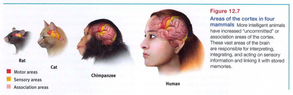
So far, we have pointed out small cortical areas that either receive sensory input or direct muscular output. Together, these occupy about one-fourth of the human brain's thin, wrinkled cover. What, then, goes on in the vast regions of the cortex? In these association areas (the peach-colored areas in FIGURE 12.7), neurons are busy with higher mental functions-many of the tasks that make us human.
Electrically probing an association area won't trigger any observable response. So, unlike the sensory and motor areas, association area functions cannot be neatly mapped. Their silence has led to what Donald McBurney (1996, p. 44) has called “one of the hardiest weeds in the garden of psychology": the claim that we ordinarily use only 10 percent of our brains. (If true, wouldn't this imply a 90 percent chance that a bullet to your brain would land in an unused area?) Surgically lesioned animals and brain-damaged humans bear witness that association areas are not dormant. Rather, these areas interpret, integrate, and act on sensory information and link it with stored memories - a very important part of thinking.
Association areas are found in all four lobes. The prefrontal cortex in the forward part of the frontal lobes enables judgment, planning, and processing of new memories. People with damaged frontal lobes may have intact memories, high scores on intelligence tests, and great cake-baking skills. Yet they would not be able to plan ahead to begin baking a cake for a birthday party (Huey et al., 2006).
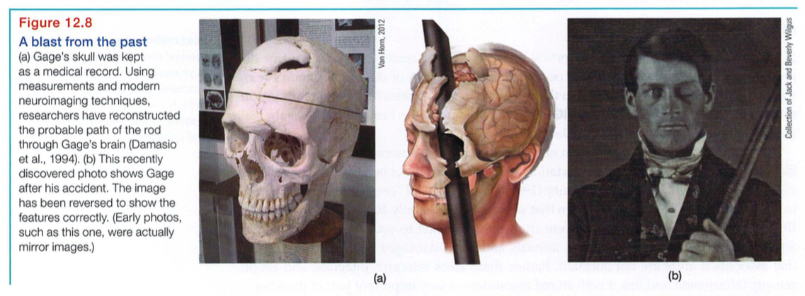
Frontal lobe damage also can alter personality and remove a person's inhibitions. Consider the classic case of railroad worker Phineas Gage. One afternoon in 1848, Gage, then 25 years old, was packing gunpowder into a rock with a tamping iron. A spark ignited the gunpowder, shooting the rod up through his left cheek and out the top of his skull, leaving his frontal lobes massively damaged (FIGURE 12.8). To everyone's amazement, he was immediately able to sit up and speak, and after the wound healed he returned to work. But the affable, soft-spoken man was now irritable, profane, and dishonest. This person, said his friends, was "no longer Gage." Although his mental abilities and memories were intact, his personality was not. (Although Gage lost his job, he did, over time, adapt to his injury and find work as a stagecoach driver [Macmillan & Lena, 2010].)
More recent studies of people with damaged frontal lobes have revealed similar impairments. Not only may they become less inhibited (without the frontal lobe brakes on their impulses), but their moral judgments may seem unrestrained by normal emotions. Would you advocate pushing someone in front of a runaway boxcar to save five others? Most people do not, but those with damage to a brain area behind the eyes often do (Koenigs et al., 2007). With their frontal lobes ruptured, people's moral compass seems to disconnect from their behavior.
Association areas also perform other mental functions. In the parietal lobes, parts of which were large and unusually shaped in Einstein's normal-weight brain, they enable mathematical and spatial reasoning (Witelson et al., 1999). In patients undergoing brain surgery, stimulation of one parietal lobe area produced a feeling of wanting to move an upper limb, the lips, or the tongue (but without any actual movement). With increased stimulation, patients falsely believed they actually had moved. Curiously, when surgeons stimulated a different association area near the motor cortex in the frontal lobes, the patients did move but had no awareness of doing so (Desmurget et al., 2009). These head-scratching findings suggest that our perception of moving flows not from the movement itself, but rather from our intention and the results we expected.
Yet another association area, on the underside of the right temporal lobe, enables us to recognize faces. If a stroke or head injury destroyed this area of your brain, you would still be able to describe facial features and to recognize someone's gender and approximate age, yet be strangely unable to identify the person as, say, Lady Gaga, or even your grandmother.
Nevertheless, we should be wary of using pictures of brain "hot spots" to create a new phrenology that locates complex functions in precise brain areas (Uttal, 2001). Complex mental functions don't reside in anyone place. There is no one spot in a rat's small association cortex that, when damaged, will obliterate its ability to learn or remember a maze.
Similarly, the acquisition, development, and use of language depends on both specialized neural networks and their integration. Nineteenth-century research by French physician Paul Broca and German investigator Carl Wernicke led to the discovery of specialized language brain areas. Damage to Broca's area disrupts speaking, while damage to Wernicke's area disrupts understanding. Today's neuroscience has shown that language functions are distributed across other brain areas as well.
Memory, language, and attention result from the synchronized activity among distinct brain areas (Knight, 2007). Ditto for religious experience. Reports of more than 40 distinct brain regions becoming active in different religious states, such as praying and meditating, indicate that there is no simple "God spot" (Fingelkurts & Fingelkurts, 2009). The big lesson: Our mental experiences arise from coordinated brain activity.
Our brains are sculpted not only by our genes but also by our experiences. MRI scans show that well-practiced pianists have a larger-than-usual auditory cortex area that encodes piano sounds (Bavelier et al., 2000; Pantev et al., 1998). In Unit IX, we'll focus more on how experience molds the brain. For now, let's turn to another aspect of the brain's plasticity: its ability to modify itself after damage.
Some of the effects of brain damage described earlier can be traced to two hard facts: (1) Severed neurons, unlike cut skin, usually do not regenerate. (If your spinal cord were severed, you would probably be permanently paralyzed.) And (2) some brain functions seem preassigned to specific areas. One newborn who suffered damage to temporal lobe facial recognition areas later remained unable to recognize faces (Farah et al., 2000). But there is good news: Some of the brain's neural tissue can reorganize in response to damage. Under the surface of our awareness, the brain is constantly changing, building new pathways as it adjusts to little mishaps and new experiences.
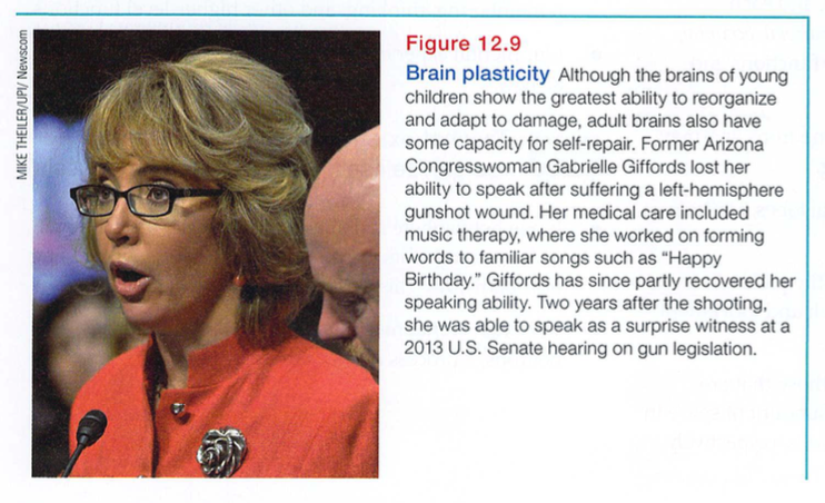
Plasticity may also occur after serious damage, especially in young children (Kolb, 1989; see also FIGURE 12.9). Constraint-induced therapy aims to rewire brains and improve the dexterity of a brain-damaged child or even an adult stroke victim (Taub, 2004). By restraining a fully functioning limb, therapists force patients to use the "bad" hand or leg, gradually reprogramming the brain. One stroke victim, a surgeon in his fifties, was put to work cleaning tables, with his good arm and hand restrained. Slowly, the bad arm recovered its skills. As damaged-brain functions migrated to other brain regions, he gradually learned to write again and even to play tennis (Doidge, 2007).
The brain's plasticity is good news for those who are blind or deaf. Blindness or deafness makes unused brain areas available for other lIses (Amedi et al., 2005). If a blind person uses one finger to read Braille, the brain area dedicated to that finger expands as the sense of touch invades the visual cortex tha t normally helps people see (Barinaga, 1992a; Sadato et al., 1996). Plasticity also helps explain why some studies find that deaf people have enhanced peripheral vision (Bosworth & Dobkins, 1999). In those people whose native language is sign, the temporal lobe area normally dedicated to hearing waits in vain for stimulation. Finally, it looks for other signals to process, such as those from the visual systenl.
Similar reassignment may occur when disease or damage frees up other brain areas normally dedicated to specific functions. If a slow-growing left hemisphere tumor disrupts language (which resides mostly in the left hemisphere), the right hemisphere may compensate (Thiel et al., 2006). If a finger is amputated, the somatosensory cortex that received its input will begin to receive input from the adjacent fingers, which then become more sensitive (Fox, 1984).
Although the brain often attempts self-repair by reorganizing existing tissue, it sometimes attempts to mend itself by producing new brain cells. This process, known as neurogenesis, has been found in adult mice, birds, monkeys, and humans Gessberger et al., 2008). These baby neurons originate deep in the brain and may then migrate elsewhere and form connections with neighboring neurons (Aimone et al., 2010; Gould, 2007) .
Master stem cells that can develop into any type of brain cell have also been discovered in the human embryo. If mass-produced in a lab and injected into a damaged brain, might neural stem cells turn themselves into replacements for lost brain cells? Might we someday be able to rebuild damaged brains, much as we reseed damaged lawns? Might new drugs spur the production of new nerve cells? Stay tuned. Today's biotech companies are hard at work on such possibilities. In the meantime, we can all benefit from other natural promoters of neurogenesis, such as exercise, sleep, and nonstressful but stimulating environments (Iso et al., 2007; Pereira et al., 2007; Stranahan et al., 2006).