

The brain enables the mind-seeing, hearing, smelling, feeling, remembering, thinking, speaking, dreaming, loving. Moreover, it is the brain that self-reflectively analyzes the brain. When we're thinking about our brain, we're thinking with our brain - by firing across millions of synapses and releasing billions of neurotransmitter molecules. Neuroscientists tell us that the mind is what the brain does. Brain, behavior, and cognition are an integrated whole. But precisely where and how are the mind's functions tied to the brain? Let's first see how scientists explore such questions.
A century ago, scientists had no tools high-powered yet gentle enough to explore the living human brain. Early case studies of patients by physicians and others helped localize some of the brain's functions. Damage to one side of the brain often caused numbness or paralysis on the body's opposite side, suggesting that the body's right side is wired to the brain's left side, and vice versa. Damage to the back of the brain disrupted vision, and to the left-front part of the brain produced speech difficulties. Gradually, these early explorers were mapping the brain.
Now, within a lifetime, a new generation of neural cartographers is probing and mapping the known universe's most amazing organ. Scientists can selectively lesion (destroy) tiny clusters of brain cells, leaving the surrounding tissue unharmed. In the laboratory, such studies have revealed, for example, that damage to one area of the hypothalamus in a rat's brain reduces eating, to the point of starvation, whereas damage in another area produces overeating.
Today's neuroscientists can also electrically, chemically, or magnetically stimulate various parts of the brain and note the effect. Depending on the stimulated brain part, people may - to name a few examples - giggle, hear voices, turn their head, feel themselves falling, or have an out-of-body experience (Selimbeyoglu & Parvizi, 2010). Scientists can even snoop on the messages of individual neurons. With tips so small they can detect the electrical pulse in a single neuron, modern microelectrodes can, for example, now detect exactly where the information goes in a cat's brain when someone strokes its whisker. Researchers can also eavesdrop on the chatter of billions of neurons and can see color representations of the brain's energy-consuming activity.
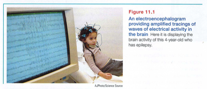
Right now, your mental activity is emitting telltale electrical, metabolic, and magnetic signals that would enable neuroscientists to observe your brain at work. Electrical activity in your brain's billions of neurons sweeps in regular waves across its surface. An electroencephalogram (EEG) is an amplified readout of such waves. Researchers record the brain waves through a shower-cap-like hat that is filled with electrodes covered with a conductive gel. Studying an EEG of the brain's activity is like studying a car engine by listening to its hum. With no direct access to the brain, researchers present a stimulus repeatedly and have a computer filter out brain activity unrelated to the stimulus. What remains is the electrical wave evoked by the stimulus (FIGURE 11.1).
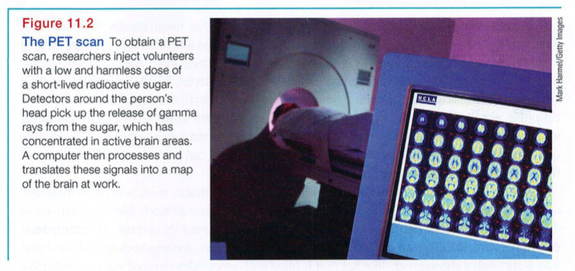
"You must look into people, as well as at them," advised Lord Chesterfield in a 1746 letter to his son. Unlike EEGs, newer neuroimaging techniques give us that Superman-like ability to see inside the living brain. For example, the CT (computed tomography) scan examines the brain by taking X-ray photographs that can reveal brain damage. Even more dramatic is the PET (positron emission tomography) scan (FIGURE 11.2), which depicts brain activity by showing each brain area's consumption of its chemical fuel, the sugar glucose. Active neurons are glucose hogs, and after a person receives temporarily radioactive glucose, the PET scan can track the gamma rays released by this "food for though t" as the person performs a given task. Rather like weather radar showing rain activity, PET-scan "hot spots" show which brain areas are most active as the person does mathematical calculations, looks at images of faces, or daydreams.
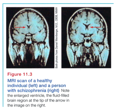
In MRI (magnetic resonance imaging) brain scans, the person's head is put in a strong magnetic field, which aligns the spinning atoms of brain molecules. Then, a radio-wave pulse momentarily disorients the atoms. When the atoms return to their normal spin, they emit signals that provide a detailed picture of soft tissues, including the brain. MRI scans have revealed a larger-than-average neural area in the left hemisphere of musicians who display perfect pitch (Schlaug et al., 1995). They have also revealed enlarged ventricles - fluid-filled brain areas (marked by the red arrows in FIGURE 11.3)-in some patients who have schizophrenia, a disabling psychological disorder.
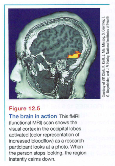
A special application of MRI - fMRI (functional MRI) - can reveal the brain's functioning as well as its structure. Where the brain is especially active, blood goes. By comparing MRI scans taken less than a second apart, researchers can watch as specific brain areas activate, showing increased oxygen-laden bloodflow. As the person looks at a scene, for example, the fMRI machine detects blood rushing to the back of the brain, which processes visual information (see Figure 12.5, in the discussion of cortex functions in Module 12).
Such snapshots of the brain's changing activity are providing new insights - albeit sometimes overstated (Vul et al., 2009a,b) - into how the brain divides its labor. A mountain of recent fMRI studies suggests which
brain areas are most active when people feel pain or rejection, listen to angry voices, think about scary things, feel happy, or become sexually excited. The technology enables a very crude sort of mind reading. After scanning 129 people's brains as they did eight different mental tasks (such as reading, gambling, or rhyming), neuroscientists were able, with 80 percent accuracy, to predict which of these mental activities people were doing (Poldrack et al., 2009). Other studies have explored brain activity associated with religious experience, though without settling the question of whether the brain is producing or perceiving God (Fingelkurts & Fingelkurts, 2009; Inzlicht et al., 2009; Kapogiannis et al., 2009) ,
* * *
Today's techniques for peering into the thinking, feeling brain are doing for psychology what the microscope did for biology and the telescope did for astronomy. From them we have learned more about the brain in the last 30 years than in the previous 30,000. To be learning about the neurosciences now is like studying world geography while Magellan was exploring the seas. This truly is the golden age of brain science,
An animal's capacities come from its brain structures. In primitive animals, such as sharks, a not-so-complex brain primarily regulates basic survival functions: breathing, resting, and feeding. In lower mammals, such as rodents, a more complex brain enables emotion and greater memory. In advanced mammals, such as humans, a brain that processes more information enables increased foresight as well.
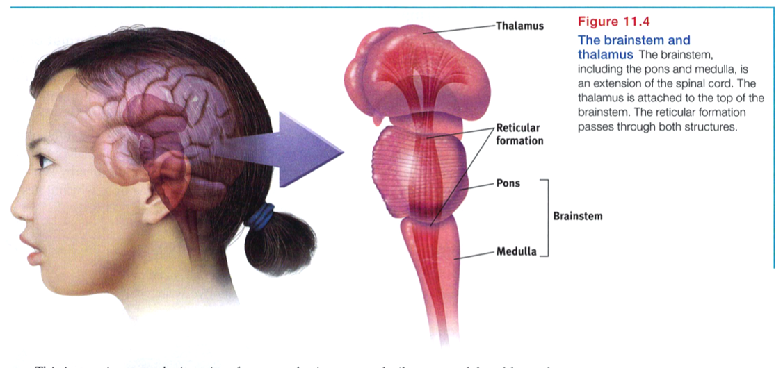
The brain's oldest and innermost region is the brainstem. It begins where the spinal cord swells slightly after entering the skull. This slight swelling is the medulla (FIGURE 11.4). Here lie the controls for your heartbeat and breathing. As some brain-damaged patients in a vegetative state illustrate, we need no higher brain or conscious mind to orchestrate our heart's pumping and lungs' breathing. The brainstem handles those tasks.
Just above the medulla sits the pons, which helps coordinate movements. If a cat's brainstem is severed from the rest of the brain above it, the animal will still breathe and live - and even run, climb, and groom (Klemm, 1990). But cut off from the brain's higher regions, it won't purposefully run or climb to get food.
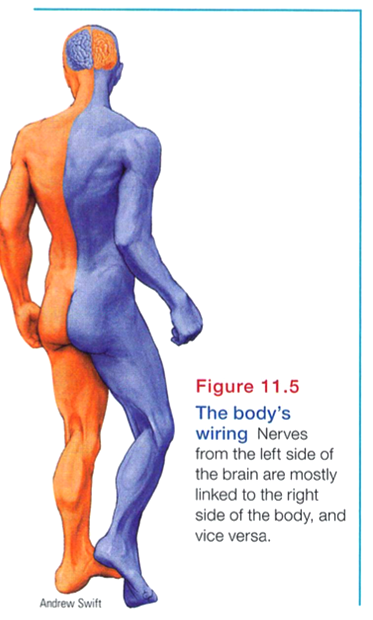
The brainstem is a crossover point, where most nerves to and from each side of the brain connect with the body's opposite side (FIGURE 11.5). This peculiar cross-wiring is but one of the brain's many surprises.
Sitting atop the brainstem is the thalamus, a pair of egg-shaped structures that act as the brain's sensory control center (Figure 11.4). The thalamus receives information from all the senses except smell and routes it to the higher brain regions that deal with seeing, hearing, tasting, and touching. The thalamus also receives some of the higher brain's replies, which it then directs to the medulla and to the cerebellum (see the next page). Think of the thalamus as being to sensory information what London is to England's trains: a hub through which traffic passes en route to various destinations.
Inside the brainstem, between your ears, lies the reticular ("netlike") formation, a neuron network that extends from the spinal cord right up through the thalamus. As the spinal cord's sensory input flows up to the thalamus, some of it travels through the reticular formation, which filters incoming stimuli and relays important information to other brain areas.
In 1949, Giuseppe Moruzzi and Horace Magoun discovered that electrically stimulating the reticular formation of a sleeping cat almost instantly produced an awake, alert animal. When Magoun severed a cat's reticular formation without damaging the nearby sensory pathways, the effect was equally dramatic: The cat lapsed into a coma from which it never awakened. The conclusion? The reticular formation enables arousal.
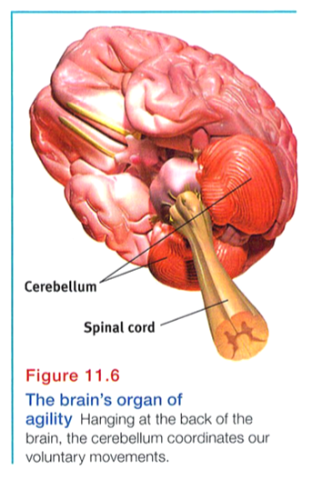
Extending from the rear of the brainstem is the baseball-sized cerebellum, meaning "little brain," which is what its two wrinkled halves resemble (FIGURE 11.6). As you will see in Module 32, the cerebellum enables nonverbal learning and memory. It also helps us judge time, modulate our emotions, and discriminate sounds and textures (Bower & Parsons, 2003). And it coordinates voluntary movement (with assistance from the pons). When a soccer player executes a perfect bicycle kick (above), give his cerebellum some credit. If you injured your cerebellum, you would have difficulty walking, keeping your balance, or shaking hands. Your movements would be jerky and exaggerated. Gone would be any dreams of being a dancer or guitarist. Under alcohol's influence on the cerebellum, coordination suffers, as many a driver has learned after being pulled over and given a roadside test.
* * *
Note: These older brain functions all occur without any conscious effort. This illustrates another of our recurring themes: Our brain processes most information outside of our awareness. We are aware of the results of our brain's labor (say, our current visual experience) but not of how we construct the visual image. Likewise, whether we are asleep or awake, our brainstem manages its life-sustaining functions, freeing our newer brain regions to think, talk, dream, or savor a memory.
What are the limbic system 's structures and functions?
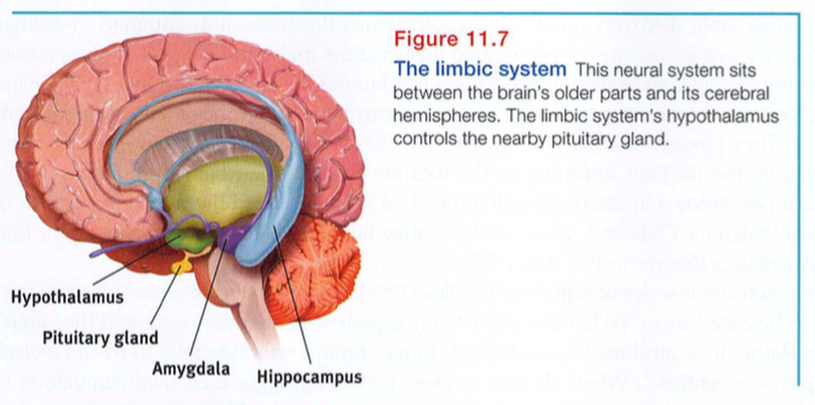
We've considered the brain's oldest parts, but we've not yet reached its newest and highest regions, the cerebral hemispheres (the two halves of the brain). Between the oldest and newest brain areas lies the limbic system (limbus means "border"). This system contains the amygdala, the hypothalamus, and the hippocampus (FIGURE 11.7). The hippocampus processes conscious memories. Animals or humans who lose their hippocampus to surgery or injury also lose their ability to form new memories of facts and events. Module 31 explams bow our two-track mind processes our memories. For now, let's look at the limbic system's links to emotions such as fear and anger, and to basic motives such as those for food and sex.
Research has linked the amygdala, two lima-bean-sized neural clusters, to aggression and fear. In 1939, psychologist Heinrich Kluver and neurosurgeon Paul Buey surgically removed a rhesus monkey's amygdala, turning the normally ill-tempered animal into the most mellow of creatures. In studies with other wild animals, including the lynx, wolverine, and wild rat, researchers noted the same effect.
What then might happen if we electrically stimulated the amygdala of a normally placid domestic animal, such as a cat? Do so in one spot and the cat prepares to attack, hissing with its back arched, its pupils dilated, its hair on end. Move the electrode only slightly within the amygdala, cage the cat with a small mouse, and now it cowers in terror.
These and other experiments have confirmed the amygdala's role in rage and fear, including the perception of these emotions and the processing of emotional memories (Anderson & Phelps, 2000; Poremba & Gabriel, 2001). But we must be careful. The brain is not neatly organized into structures that correspond to our behavior categories. When we feel or act in aggressive or fearful ways, there is neural activity in many levels of our brain. Even within the limbic system, stimulating structures other than the amygdala can evoke aggression or fear. If you charge your cell phone's dead battery, you can activate the phone and make a call. Yet the battery is merely one link in an integrated system.
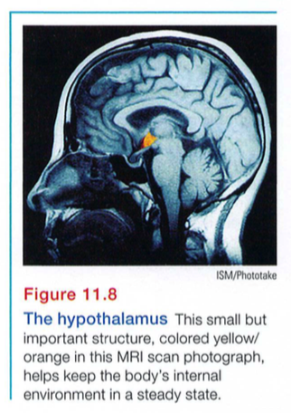
Just below (hypo) the thalamus is the hypothalamus (FIGURE 11.8), an important link in the command chain governing bodily maintenance. Some neural clusters in the hypothalamus influence hunger; others regulate thirst, body temperature, and sexual behavior. Together, they help maintain a steady internal state. As the hypothalamus monitors the state of your body, it tunes into your blood chemistry and any incoming orders from other brain parts. For example, picking up signals from your brain's cerebral cortex that you are thinking about sex, your hypothalamus will secrete hormones. These hormones will in turn trigger the adjacent "master gland," your pituitary (see Figure 11.7), to influence your sex glands to release their hormones. These will intensify the thoughts of sex in your cerebral cortex. (Once again, we see the interplay between the nervous and endocrine systems: The brain influences the endocrine system, which in turn influences the brain.)
A remarkable discovery about the hypothalamus illustrates how progress in science often occurs - when curious, open-minded investigators make an unexpected observation. Two young McGill University neuropsychologists, James Olds and Peter Milner (1954), were trying to implant an electrode in a rat's reticular formation when they made a magnificent mistake: They placed the electrode incorrectly (Olds, 1975). Curiously, as if seeking more stimulation, the rat kept returning to the location where it had been stimulated by this misplaced electrode. On discovering that they had actually placed the device in a region of the hypothalamus, Olds and Milner realized they had stumbled upon a brain center that provides pleasurable rewards (Olds, 1975).
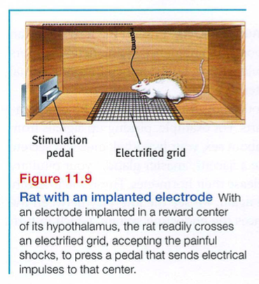
In a meticulous series of experiments, Olds (1958) went on to locate other "pleasure centers," as he called them. (What the rats actually experience only they know, and they aren't telling. Rather than attribute human feelings to rats, today's scientists refer to reward centers, not "pleasure centers.") When allowed to press pedals to trigger their own stimulation in these areas, rats would sometimes do so at a feverish pace-up to 7000 times per hour-until they dropped from exhaustion. Moreover, to get this stimulation, they would even cross an electrified floor that a starving rat would not cross to reach food (FIGURE 11.9).
Other limbic system reward centers, such as the nucleus accumbens in front of the hypothalamus, were later discovered in many other species, including dolphins and monkeys. In fact, animal research has revealed both a general dopamine-related reward system and specific centers associated with the pleasures of eating, drinking, and sex. Animals, it seems, come equipped with built-in systems that reward activities essential to survival.
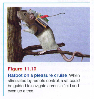
Contemporary researchers are experimenting with new ways of using limbic stimulation to control animals' actions in future applications, such as search -and -rescue operations. By rewarding rats for turning left or right, one research team trained previously caged rats to navigate natural environments (Talwar et al., 2002; FIGURE 11.10). By pressing buttons on a laptop, the researchers were then able to direct the rat - which carried a receiver, power source, and video camera on a backpack - to turn on cue, climb trees, scurry along branches, and turn around and come back down.
Do humans have limbic centers for pleasure? Indeed we do. To calm violent patients, one neurosurgeon implanted electrodes in such areas. Stimulated patients reported mild pleasure; unlike Olds' rats, however, they were not driven to a frenzy (Deutsch, 1972; Hooper & Teresi, 1986).
Experiments have also revealed the effects of a dopamine-related reward system in people. One research team had people rate the desirability of different vacation destinations. Then, after receiving either a dopamine-increasing drug or a sugar pill, they imagined themselves vacationing at half the locations. A day later, when presented with pairs of vacation spots they had initially rated equally, only the dopamine takers preferred the places they had imagined under dopamine's influence (Sharot et al., 2009) . The participants, it seems, associated the imagined experiences with dopamine-induced pleasant feelings.
Some researchers believe that addictive disorders, such as substance use disorders and binge eating, may stem from malfunctions in natural brain systems for pleasure and wellbeing. People genetically predisposed to this reward deficiency syndrome may crave whatever provides that missing pleasure or relieves negative feelings (Blum et a1., 1996).
* * *